Tooth loss affects millions of adults worldwide, stemming from factors like severe decay, periodontal disease, traumatic injury, or congenital conditions. Beyond the obvious aesthetic changes, missing teeth lead to functional challenges, such as difficulty chewing, impaired speech, and progressive bone loss in the jaw. When a tooth root is no longer present to stimulate the surrounding jawbone, the bone tissue naturally resorbs over time, altering facial structure and jeopardizing the stability of neighboring teeth.
To address both structural and cosmetic concerns, restorative dentistry relies on permanent tooth replacement solutions. Patient education resources from Hughson Dental Implants explain how modern implant fixtures replicate the natural root structure, stimulating bone growth and preventing the facial collapse associated with untreated tooth loss. Understanding the biological mechanisms, surgical steps, and long-term care required for dental implants empowers patients to make informed decisions about their oral health.
The Biological Foundation: What Is Osseointegration?
The success of a dental implant relies on a unique biological process called osseointegration. Unlike traditional removable dentures or fixed bridges—which sit on top of the gums or rely on adjacent teeth for support—a dental implant integrates directly with the jawbone.
The Material Science Behind Implants
Implants are primarily crafted from biocompatible materials, most commonly medical-grade titanium or zirconia.
- Titanium: Renowned for its exceptional strength, durability, and biological compatibility. Human bone cells naturally attach to titanium surfaces without treating the metal as a foreign object.
- Zirconia: A high-strength ceramic alternative that offers a tooth-colored, metal-free option for patients with metal sensitivities or thin gum tissue where dark metal might show through.
How Bone Fuses to the Post
Once surgically inserted into the jawbone, the implant fixture acts as an artificial tooth root. Over a period of three to six months, osteoblast cells build new bone matrix directly around the micro-textured surface of the post. This structural fusion creates a stable foundation capable of withstanding heavy bite forces.
Anatomy of a Dental Implant System
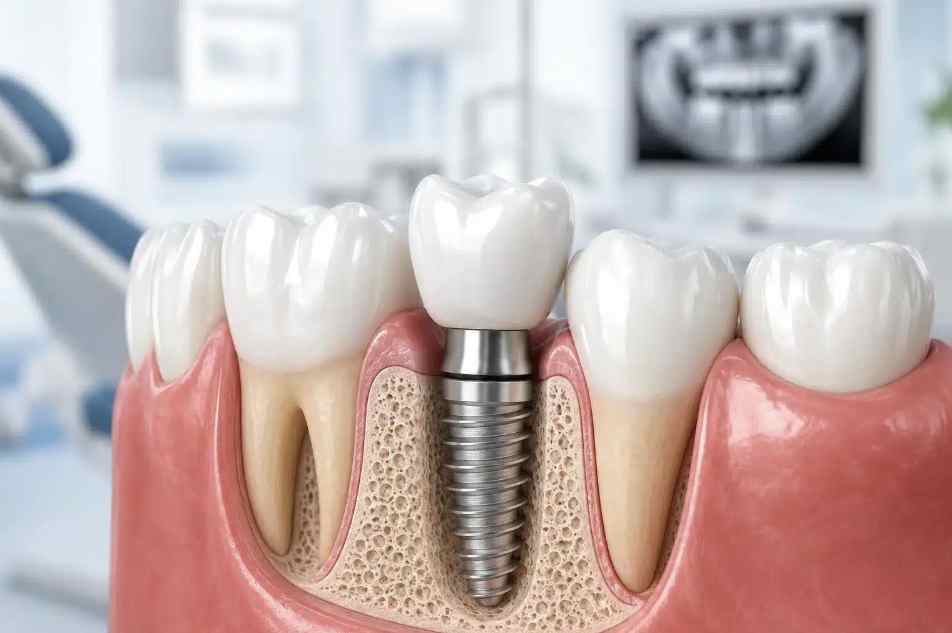
A standard dental implant restoration consists of three distinct components:
- The Implant Post: A small, threaded screw surgically embedded into the jawbone beneath the gumline.
- The Abutment: A connecting piece attached to the top of the post once osseointegration is complete. It extends through the gumline to hold the final restoration.
- The Prosthetic Crown: The visible, custom-crafted artificial tooth made from porcelain or ceramic that matches the shape, size, and color of surrounding natural teeth.
The Step-by-Step Implant Process
Receiving a dental implant is a multi-step procedure that unfolds over several months to allow proper healing at each stage.
┌──────────────────────────────────────────────────────────┐
│ Dental Implant Care Protocol │
├──────────────────────────┬───────────────────────────────┤
│ Evaluation Phase │ 3D CBCT Scans & Bone Density │
│ │ Mapping │
├──────────────────────────┼───────────────────────────────┤
│ Surgical Placement │ Implant Post Insertion into │
│ │ Jawbone │
├──────────────────────────┼───────────────────────────────┤
│ Osseointegration Period │ 3-6 Months of Bone Fusion │
│ │ & Healing │
├──────────────────────────┼───────────────────────────────┤
│ Restoration Stage │ Abutment Placement & Custom │
│ │ Crown Attachment │
└──────────────────────────┴───────────────────────────────┘
Phase 1: Comprehensive Initial Assessment
Before surgery, a thorough clinical exam is performed using 3D Cone Beam Computed Tomography (CBCT) scanning. This detailed imaging evaluates jawbone density, maps nerve pathways, and locates sinus cavities to ensure safe, precise placement.
Phase 2: Bone Grafting (If Necessary)
If a patient has experienced significant bone loss due to prolonged tooth absence, a bone graft may be needed. Bone graft material is placed in the depleted area to build sufficient bone volume, establishing a sturdy base for the implant post.
Phase 3: Surgical Placement
Under local anesthesia or conscious sedation, the clinician creates a small incision in the gum tissue, prepares a precise channel in the jawbone, and threads the titanium post into place. The gum tissue is then stitched closed over or around the fixture.
Phase 4: Healing and Osseointegration
During the healing phase, patients may wear a temporary cosmetic restoration. It is critical to avoid placing heavy biting pressure on the implant area while bone cells actively fuse with the post surface.
Phase 5: Abutment Placement and Final Restoration
Once integration is verified via X-rays, the top of the post is exposed, and the abutment is attached. Impressions are taken to craft a custom porcelain crown, which is permanently cemented or screwed onto the abutment to finish the restoration.
Comparing Tooth Replacement Options
| Feature | Dental Implant | Fixed Dental Bridge | Removable Denture |
| Jawbone Preservation | Yes (Stimulates bone growth) | No (Bone resorbs over time) | No (Bone resorbs over time) |
| Impact on Neighboring Teeth | None (Standalone unit) | High (Requires reshaping adjacent teeth) | Low to Moderate (Rests on existing teeth) |
| Lifespan | 20+ Years to Lifetime | 7 to 15 Years | 5 to 7 Years |
| Maintenance | Normal brushing and flossing | Specialized flossing underneath | Daily removal and soaking |
Essential Daily Care for Implant Longevity
Although dental implants cannot develop tooth decay like natural teeth, the surrounding gum tissue can still become inflamed or infected if plaque accumulates.
- Perform Thorough Plaque Removal: Brush twice daily using a soft-bristled toothbrush or an low-abrasive toothpaste to protect the crown surface.
- Clean Interdental Spaces Daily: Use woven dental floss, interdental brushes, or a water flosser to clean thoroughly around the abutment and under the crown margin.
- Prevent Peri-Implantitis: Peri-implantitis is an inflammatory condition affecting the soft and hard tissues around an implant. Poor oral hygiene allows bacteria to accumulate, potentially causing bone loss around the post and leading to implant failure.
- Schedule Regular Clinical Audits: Visit your dentist every six months for professional cleanings and X-rays to monitor bone stability around the fixture.
Understanding the biology of osseointegration and following a consistent oral hygiene routine allows individuals to replace missing teeth effectively, protecting their jawbone structure and maintaining a healthy, functional smile for decades.